Science
Researchers Achieve Breakthrough in Cancer Treatment with Radioactive Ion Beams
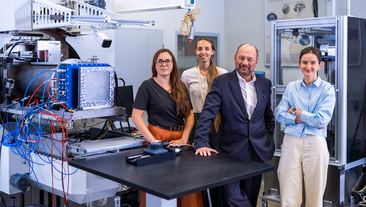
Researchers in Germany have successfully conducted the first cancer treatment using a radioactive carbon ion beam, specifically 11C, on a mouse with a bone tumour located near the spine. This innovative approach, part of the BARB project funded by the European Research Council, leverages radioactive ion beams for simultaneous treatment and imaging, marking a significant step forward in particle therapy.
Particle therapy, which utilizes beams of protons or heavy ions, has gained recognition for its effectiveness in targeting tumours with precision. The technique exploits the Bragg peak phenomenon, allowing for highly conformal dose deposition. However, this precision also makes the treatment sensitive to range uncertainties, which can adversely affect the positioning of the Bragg peak. Researchers aim to mitigate these uncertainties by employing positron emission tomography (PET) to map isotopes generated during the treatment as they interact with the patient’s tissues.
Currently, particle therapy with carbon ions, which is available at 17 centres worldwide, relies on detecting beta decay signals from 10C and 11C fragments. The challenge lies in the fact that these fragments produce a weak PET signal and their lower mass can shift the measured activity peak away from the Bragg peak.
According to Marco Durante, head of biophysics at the GSI Helmholtz Centre for Heavy Ion Research and principal investigator of the BARB project, using positron-emitting ions like 11C could provide a solution. “Range uncertainty remains the main problem of particle therapy, as we do not know exactly where the Bragg peak is,” Durante explained. He emphasized that combining a radioactive beam with PET imaging allows for real-time localization and adjustments during treatment, thereby enhancing the precision of the therapy.
Innovative Experimental Approach
To validate this concept, Durante and his team conducted in vivo experiments at the GSI/FAIR accelerator facility in Darmstadt. They utilized a portable small-animal in-beam PET scanner developed by Katia Parodi and her team at LMU Munich. This sophisticated scanner, designed for the ERC project SIRMIO, features 56 depth-of-interaction detectors made of pixelated LYSO crystals, offering exceptional sensitivity and spatial resolution.
During the experiments, the researchers treated 32 mice with osteosarcoma tumours implanted near the spinal cord using a radioactive 11C ion beam produced at the GSI fragment separator. To ensure comprehensive coverage of the target volume, they employed a range modulator to create a spread-out Bragg peak (SOBP) and a plastic compensator collar for positioning and immobilizing the mice. The animals were treated under anaesthesia, receiving either 20 Gy or 5 Gy at a rate of approximately 1 Gy/min.
The team meticulously compared the measured activity with simulations based on pre-treatment microCT scans. Notably, they observed a shift in activity distributions of about 1 mm, attributed to anatomical changes between the scans and the treatment positioning. After adjusting for this shift, the simulation aligned closely with the actual measurements. “Our findings reinforce the necessity of vertical CT planning and highlight the potential of online PET as a valuable tool for upright particle therapy,” the researchers noted.
Despite the close proximity of the tumour to the spinal cord, which raises concerns about potential damage, the online PET images confirmed that the SOBP did not impact the spine. The team monitored the mice for any signs of radiation-induced myelopathy, a condition that can lead to motor deficits or paralysis, and found no severe toxicity, indicating that the spinal cord was not exposed to harmful doses.
Future Prospects and Clinical Translation
Following the treatment, the team assessed tumour responses, revealing complete tumour control after 20 Gy irradiation and significant growth delay after 5 Gy. These results suggest effective target coverage across all subjects. Moreover, the researchers evaluated the washout of the PET signal from the tumour, which exhibited a dose-dependent decay pattern. Durante speculated that the high-dose radiotherapy might work differently than conventional methods by damaging blood vessels supplying the tumour, thereby effectively “starving” it.
Looking ahead, the research team plans to explore the use of 10C or 15O treatment beams, which are expected to produce stronger signals and enhanced temporal resolution. The new Super-FRS fragment separator at the FAIR accelerator facility is anticipated to provide the high-intensity beams necessary for these studies.
For clinical translation, Durante acknowledged the need for a feasible and cost-effective design. He mentioned ongoing efforts at CERN, specifically the MEDICIS-Promed project, which aims to create a design based on isotope separation online (ISOL) for use in current accelerators. Furthermore, GSI is investigating the potential for an in-flight device for medical accelerators.
The findings from this research have been published in Nature Physics, underscoring the significant advancements in cancer treatment methodologies through the application of radioactive ion beams.

NASA Investigates Speedy Object 3I/ATLAS, Sparking Speculation

Reading FC Dismisses Manager Noel Hunt After Tough Loss

Ireland Offers £72,240 Grants to Attract New Residents to Islands

Niksic Earns Title of European Capital of Culture for 2030

Lidl Announces Plans for New Supermarkets in Powys by 2025

Adriatic Film & TV Awards Recognize Top Talent in Tivat

Mid Ulster Council Chair Expresses Sympathy for Deputy’s Loss

Lottery Win Turns Sour: Couple Regrets Life-Altering Jackpot

Kerry Katona and Boyfriend Share Affectionate Moments in Manchester

Neurologist Warns Excessive Use of Supplements Can Harm Brain

Fiona Phillips’ Husband Shares Heartfelt Update on Her Alzheimer’s Journey

Brian Cox Addresses Claims of Alien Probe in 3I/ATLAS Discovery

NASA Investigates Unusual Comet 3I/ATLAS; New Findings Emerge

Scientists Examine 3I/ATLAS: Alien Artifact or Cosmic Oddity?

Cole Palmer’s Cryptic Message to Kobbie Mainoo Following Loan Talks

Kerry Katona Discusses Future Baby Plans and Brian McFadden’s Wedding

Emmerdale Faces Tension as Dylan and April’s Lives Hang in the Balance

Love Island Star Toni Laite’s Mother Expresses Disappointment Over Coupling Decision
-
 Health2 months ago
Neurologist Warns Excessive Use of Supplements Can Harm Brain
-
 Health2 months ago
Fiona Phillips’ Husband Shares Heartfelt Update on Her Alzheimer’s Journey
-
 Science7 days ago
Brian Cox Addresses Claims of Alien Probe in 3I/ATLAS Discovery
-
 Science5 days ago
NASA Investigates Unusual Comet 3I/ATLAS; New Findings Emerge
-
 Science2 days ago
Scientists Examine 3I/ATLAS: Alien Artifact or Cosmic Oddity?
-
 World2 months ago
Cole Palmer’s Cryptic Message to Kobbie Mainoo Following Loan Talks
-
 Entertainment3 months ago
Kerry Katona Discusses Future Baby Plans and Brian McFadden’s Wedding
-
 Entertainment3 months ago
Emmerdale Faces Tension as Dylan and April’s Lives Hang in the Balance
-
 Entertainment3 months ago
Love Island Star Toni Laite’s Mother Expresses Disappointment Over Coupling Decision
-

 Entertainment2 months ago
Major Cast Changes at Coronation Street: Exits and Returns in 2025
-

 World2 months ago
Coronation Street’s Asha Alahan Faces Heartbreaking Assault
-

 Entertainment2 weeks ago
Stefan Dennis and Dianne Buswell Share Health Update on Strictly Come Dancing